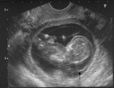

This underlines the importance of ultrasound as a noninvasive screening tool for foetal abnormalities and associated underlying trisomies in younger women (Dicke and Crane, 1991). Overall 80% of children with a chromosomal abnormality are born to women under age 35 (Savva et al., 2010). The relationship of trisomy 13 and 18 with maternal aging is, contrary to trisomy 21, less outspoken. Of those who alive at birth, only 20% will survive the first month of life and only 5% will survive the first six months (Fleischer et al., 2011). Most foetuses with trisomy 13 die in utero or are stillborn. Trisomy 13, or Patau syndrome, has a prevalence of births. Approximately 68% of foetuses with trisomy 18 die in utero, only 10% survive the first year of life (Dicke and Crane, 1991). Trisomy 18, also known as Edwards syndrome, is the second most common autosomal trisomy with a prevalence ranging between 1:3500 and 1:8000 births. Although less prevalent than trisomy 21 (1 per 800 births), they are by no means extremely rare (Bundy et al., 1986 Shields et al., 1998 Fleischer et al., 2011). Trisomy 13 as well as trisomy 18 are typified by various foetal malformations. Mostly the foetal pathology correlated with the sonographic evaluation. Most of the ultrasound features were predominant in foetuses with trisomy 18. In most cases the sonographic signs correlated with the pathology findings.Ĭonclusion: Trisomy 13 as well as trisomy 18 are characterized by a number of various malformations in the foetus. Most malformations were predominant in trisomy 18, except for genitourinary tract anomalies. The most common malformations associated with trisomy 18 were limb abnormalities and intrauterine growth restriction. Results: In foetuses with trisomy 13 the most common malformations were craniofacial defects, cerebral malformations and genitourinary tract anomalies. We evaluated the predominant signs leading to referral, the difference and overlap in presenting signs between both syndromes and when the data were available, we also compared the echographic signs with the foetal pathology.

47 Of these cases were foetuses with trisomy 13 or trisomy 18. Materials and Methods: In a period of thirty years, 1110 cases of foetal malformations were detected during specialized echographic evaluation. Objective: To determine and list the variety of the predominant appeal signs leading to referral and their accompanying features found during specialized ultrasound evaluation in foetuses with trisomy 13 and trisomy 18.


 0 kommentar(er)
